Tomographie par Cohérence Ophtique OCT NIDEK
RS-3000
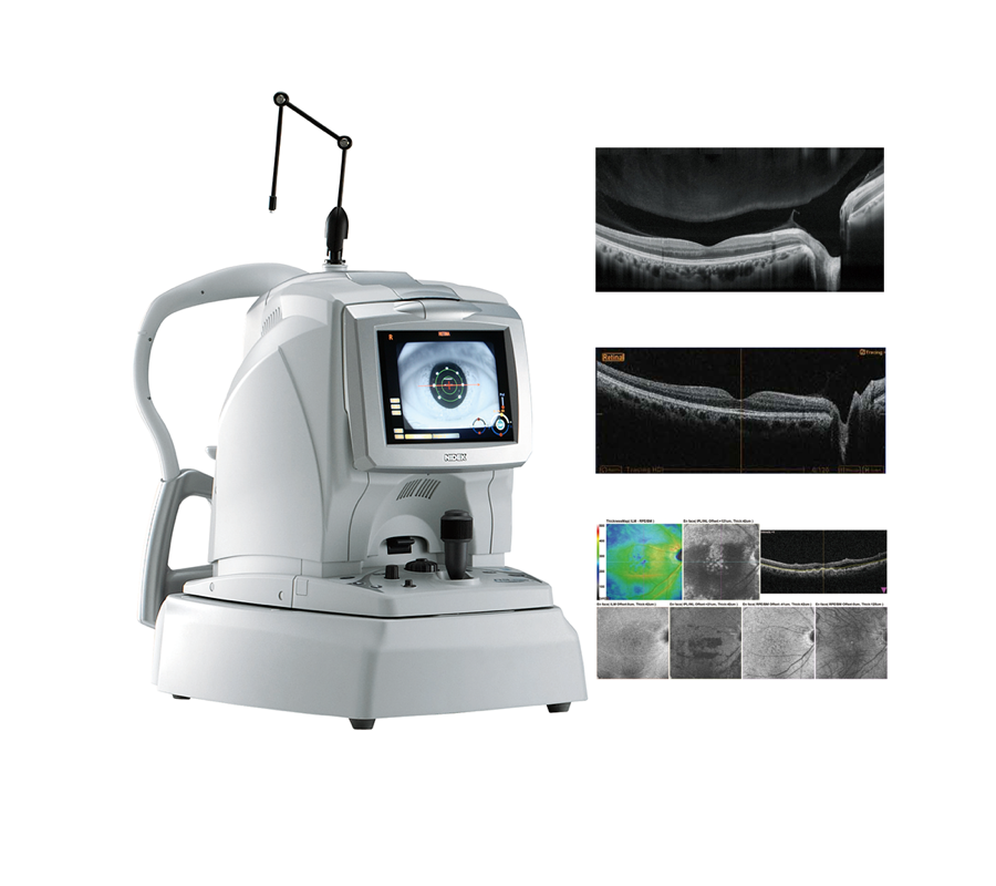
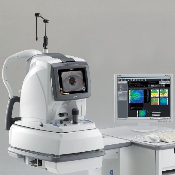
The RS-3000 Advance is a premier OCT system incorporating Scanning Laser Ophthalmoscope that is designed for comprehensive evaluation of the retina and choroid. The RS-3000 Advance provides exquisite detail of the retinal and choroidal microstructures to assist in clinical diagnosis.
Key Features:
• Wide area scan (9 x 9 mm) and high-definition OCT with SLO tracing
• Tracing HD
• High speed scanning – 53,000 A-scans/sec
• Selectable OCT sensitvity
• Torsion Eye Tracer (TET)
Wide Area and High Definition OCT with SLO Tracing
12 mm wide horizontal scan available with the RS-3000 Advance allows detailed observation of the vitreous body, retina, and choroid from the macula to optic disc in a single image.
Selectable OCT Sensitivity
Selecting the OCT sensitivity based on ocular pathology allows image capture with higher definition or at high speed. Ultra-fine, fine, and regular sensitivities are available for the RS-3000 Advance. Ultra-fine and fine sensitivities are used to capture high definition images and regular is used to capture images at high speed.
Tracing HD Plus
The tracing HD plus function in the RS-3000 Advance traces involuntary eye movements to maintain the same scan location on the SLO image for accurate image capture. This function allows accurate averaging of up to 120 images.
- Macula multi (cross)
The macula multi scan pattern captures 5 cross-sectional images each in the X and Y directions. High-quality images are easily obtained with the tracing HD plus function.
- Macula radial and disc radial
The macula radial and disc radial scan patterns capture 6 or 12 radial cross-sectional images centered on the macula and optic disc respectively. The tracing HD plus function ensures
the scan is centered on the targeted region.
- HD checker
The HD checker function in the RS-3000 Advance displays the image during averaging and allows an operator to check and finish capturing prior to reaching the number for averaging set
by an operator if sufficient image quality is obtained.
Torsion Eye Tracer (TET)
The TET incorporated in the RS-3000 Advance ensres accurate image capture by utilizing fundus information form the high definition SLO image. The three functions, positioning, tracing, and auto shot allow accurate image capture of the targeted region. Ocular cyclotorsion is traced via the torsion correction feature added to the tracing function.
Torsion correction
The torsion correction function ensures the scan is oriented at the right angle even in cases of ocular cyclotorsion and fundus tilt due to head movement or incorrect positioning of the chinrest and forehead rest.
Follow-up Image Capture
The follow-up image capture function in the RS-3000 Advance performs positioning based on the previously captured baseline data, and then tracing and auto shot. It provides ease-of-use and high reproducibility of the image capture for follow-up examination.