Echographe cardiaque portable
Orcheo Lite CV
Orcheo Lite CV - created especially for cardiovascular researches.
Orcheo Lite CV unites everything the image mode: from technology of a fabric harmonica (THI) before Doppler visualization of fabrics (TDI), a stress - an echo, cardiocalculations, a constant and wave dopler and others.
With Orcheo Lite CV you do not need to select between quality the images and an ortativnost any more.
Its interface easy in use lifts quality of ultrasonic diagnostics on new level and allows you to do your work quicker, more safely and more effectively.
Exclusive programs of measurements and the built-in data of survey of patients do Orcheo Lite CV by the ideal device for clinical trials.
Thanks to the open configuration Orcheo Lite CV can be improved with ease. You the first learn about technologies of the future and will be able to use them in the practice.
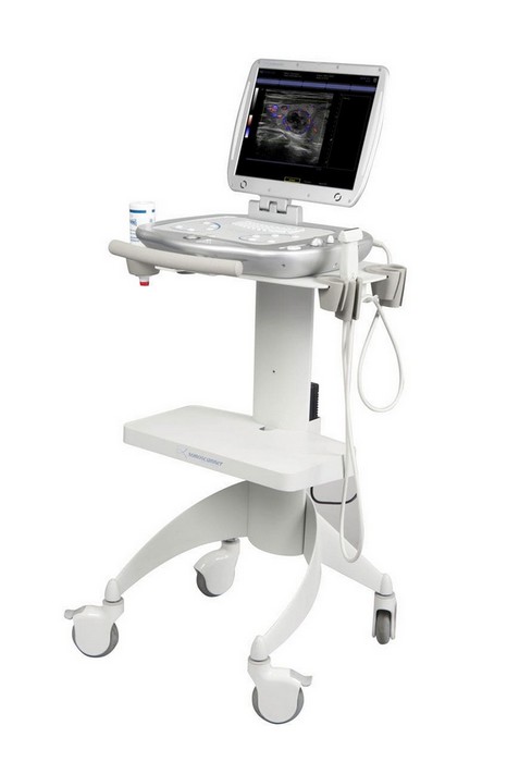
Portable perfection
OrcheoLite CV weighs only 5 kg is the real scanner of the class hand-carry that will allow carries out high-quality diagnostics in any place and at any time.
OrcheoLite CV has the high-performance battery of the longest action in the world. This integrated battery allows OrcheoLite to work continuously an hour and a half in the most extreme conditions of ambulance or sports medicine.
Large-format 15" the liquid crystal (LCD) monitor with high resolution allows to receive the accurate image.
OrcheoLite CV has huge memory size - 250 GB of which more, than it is enough, for record and storage bolshogokolichestvadanny. Such capacity of the memory device is especially useful, to record of crude data for post-processing.
Clinical range of researches
• Cardiology
• Research of vessels
• Intraoperative
• Transkranialny researches
• Small and superficial bodies
• Mammary gland
• Pediatrics and neonatology
• Research of vessels
• Abdominal researches
• Obstetrics and gynecology (4D)
• Researches of bone and muscular system
• Urology
• Biopsy
Full range of ways of the image
• B-mode
• M-mode
• Color doppler
• Pulse and wave dopler
• Constant and wave dopler
• The duplex itripleksny mode in real time with automatic trace and calculation of parameters of a Doppler range
• The power and directed power doppler - provide excellent permission with the improved color sensitivity. It is especially valuable to research of deeply located bodies and thin vessels when special accuracy is required
• High-frequency repetition of impulses (HPRF) - prevents emergence of a shumovn of the image when research to be conducted at high frequencies.
• Automatic optimization of the image Autoset - allows the user simple pressing of the button automatically to optimize the image in the 2D and spectral Doppler mode, thus avoiding tiresome manual adjustment of parameters.
• Fabric dopler
• Anatomic M-mode
The iSteer technology – scans the studied area under different corners, for improvement of visualization of tissues of relatives on density, at research of musculoskeletal system, also provides high-quality visualization when carrying out a biopsy.
The fabric harmonica - is based on technology of the second harmonica, combines ability of deep penetration of an ultrasonic beam with the fine permission and the contrast image. The technology is recommended for survey of "difficult" patients.
Trapezoid scanning – allows to expand considerably an image field, a dlyapolucheniye of fuller information on the studied body.
Technology 3D/4D
OrcheoLite CV - the real break in ultrasound as it - the first compact decision for 3D/4D technologies is valid, allows to receive the image in real time and in the mode offline.
• Exact anatomic image: use of spetsialnykhmekhanichesky sensors for transfer of the image 3D in real time and the mode offline allows the user to see the volume image with exact anatomic izobrazheniyem
• The mode 4D allows to see with a clearness the three-dimensional image of object in the movement.
• Management 3D/4D of the mode is available both in the survey mode, and after survey.
• A large number function for obstetrics and gynecology.
• 3D/4D allows you to construct the image easily and quickly.
Unique own developments of Sonoscanner
ERIOS: Extended Raw Image Optimization Station
The ERIOS technology - opens ample opportunities in post-processing of the image in real time, the frozen image, and also the kept images and video clips, allows to take repeated measurements and calculations, to operate sensitivity and contrast, dynamic range, to smooth the image for achievement of the maximum quality of the image and for the profound analysis
The RTAS-technology optimum smoothing of the image, one of the most often used tools in ERIOS. Allows to suppress effectively noise without resolution loss. As a result you receive the image which was more detailed for fast and exact statement of the diagnosis.